
Peer Reviewed
An Atlas of Lingual Lesions, Part 4
Hemangioma
Hemangiomas are benign vascular tumors due to proliferation of endothelial cells.1 Characteristically, hemangiomas are not clinically apparent at birth.2 They usually appear in the first few weeks of life as areas of pallor, followed by telangiectatic patches.2 These lesions are characterized by a distinctive life cycle in which proliferation is generally limited to the first year of life, at which time the growth rate slows to parallel the growth of the child, followed by a variable involution phase.2-5
Typically, hemangiomas are asymptomatic.2 Superficial lesions are bright red, protuberant, and sharply demarcated and are often referred to as “strawberry hemangiomas” or “capillary hemangiomas.”2 Deep lesions are bluish and dome-shaped and are noted later than superficial hemangiomas.2 They reach their maximum size between 1 and 2 years of age.2 Deep hemangiomas consists of deep, irregular, large, thin-walled cavernous vessels and sinusoids lined by epithelial cells and separated by a scanty connective tissue stroma.1,3 They feel like a “bag of worms” and are compressible; they are often referred to as “cavernous hemangiomas.”6 Approximately 60% of hemangiomas are superficial, 15% are deep, and 25% are mixed superficial and deep.6 Mixed hemangiomas (both superficial and deep) may show characteristic features of both, often presenting with a red plaque overlying a bluish nodule.2
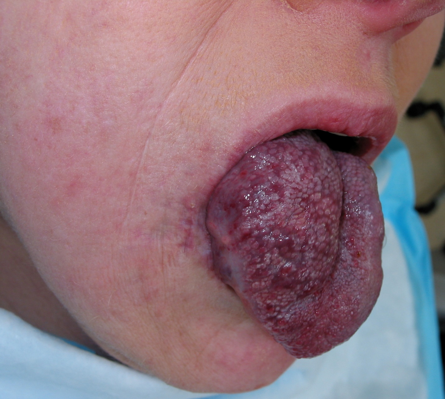
Although hemangiomas can appear anywhere on the skin, internal organs, or mucous membranes, 60% to 70% of cases occur in the head and neck region.3 However, occurrence within the oral cavity, in particular the tongue, is exceeding rare.3-7 Typically, oral hemangiomas present at an older age than lesions elsewhere, usually between the second and fourth decade.8 Most lingual hemangiomas are cavernous or mixed in nature and often present as a smooth, lobulated mass or as macroglossia (Figure).5,7 They are dark red-blue or deep red in color and soft to firm in consistency.7,8
A lingual hemangioma must be differentiated from a venolymphatic malformation, arteriovenous malformation, and lymphangioma. In the case of the patient shown in the Figure, color Doppler ultrasonography of the tongue showed a huge echogenic lesion with intermittent color picking, suggestive of a vascular lesion, and small cystic areas and increased vascularity, confirming the diagnosis of hemangioma causing macroglossia.
In the white population, hemangioma affects approximately 1.1% to 2.6% of newborn infants and 10% to 12% of children by the first year of life.9 The female to male ratio is approximately 3 to 1.4
Hemangiomas arise from endothelial stem cells that later proliferate by vasculogenesis, with further angiogenesis.2 Hypoxia and estrogen are important stimuli and have a synergistic effect on angiogenesis.10 The genes encoding vascular endothelial growth factor, indoleamine 2,3-dioxygenase, insulinlike growth factor 2, angiopoietin 1, angiopoietin 2, basic fibroblast growth factor, Cip1-interacting zinc finger protein 1, and tyrosine-protein kinase receptor (Tie2) are believed to play a significant role in the pathogenesis of hemangiomas.3,9,10
Complications include cosmetic disfigurement, recurrent trauma due to biting of the tongue, hemorrhage, ulceration, infection in an ulcerated lesion, chewing/swallowing/feeding difficulties, speech impairment, and airway obstruction.1,3,11-13 In general, the risk of complications is closely related to the size of the lesion.
Treatment should be individualized. Most lingual hemangiomas, especially small ones, may not require intervention except reassurance and watchful observation.1,3 Indications for active intervention include severe or recurrent hemorrhage, life- or function-threatening complications, pedunculated hemangiomas, and significant disfigurement.2 When treatment is necessary, surgical excision is the treatment of choice when feasible. Other treatment options include intralesional corticosteroids, oral β-blockers, sclerotherapy, laser surgery, laser photocoagulation, electrocautery, cryotherapy, immunomodulatory therapy with interferon alfa-2a, embolization, radiofrequency tissue ablation, and ligation.1,3,4,14,15
REFERENCES:
- Portaro S, Naro A, Guarneri C, Di Toro G, Manuli A, Calabrò RS. Hemangiomas of the tongue and the oral cavity in a myotonic dystrophy type 1 patient: a case report. Medicine (Baltimore). 2018;97(48):e13448.
- Leung AKC, Barankin B, Hon KL. Infantile hemangioma. Pediatr Neonatal Nurs Open J. 2014;1(1):1-6.
- Kamala KA, Ashok L, Sujatha GP. Cavernous hemangioma of the tongue: a rare case report. Contemp Clin Dent. 2014;5(1):95-98.
- V P, Puppala N, Deshmukh SN, Jagadesh B, Anuradha S. Cavernous hemangioma of tongue: management of two cases. J Clin Diagn Res. 2014;8(10):ZD15-ZD17.
- Shrestha AL, Paudel SB. Lingual cavernous hemangioma in a Nepalese boy—‘a difficult associate!!!’ J Surg Case Rep. 2018;2018(10):rjy283.
- Atherton DJ. Infantile hemangiomas. Early Hum Dev. 2006;82(12):789-795.
- Kripal K, Rajan S, Ropak B, Jayanti I. Cavernous hemangioma of the tongue. Case Rep Dent. 2013;2013:898692.
- Tasker LJ, Geoghegan J. Giant cavernous haemangioma of the tongue. Anaesthesia. 2005;60(10):1043.
- Lo K, Mihm M, Fay A. Current theories on the pathogenesis of infantile hemangioma. Semin Ophthalmol. 2009;24(3):172-177.
- Chen TS, Eichenfield LF, Friedlander SF. Infantile hemangioma: an update on pathogenesis and therapy. Pediatrics. 2013;131(1):99-108.
- Wang Y, Li X, Zhang J, et al. CIZ1 expression is upregulated in hemangioma of the tongue [published online November 19, 2018. Pathol Oncol Res. doi:10.1007/s12253-018-0495-4.
- 12. Gallarreta FW, Pieroni KA, Mantovani CP, Silva FW, Nelson-Filho P, de Queiroz AM. Oral changes stemming from hemangioma of the tongue. Pediatr Dent. 2013;35(2):E75-E78.
- Rudingwa P, Sundararajan V, Vasudevan A, Pannerselvam S. An innovative airway management technique in an infant with tongue hemangioma. Paediatr Anaesth. 2019;29(4):389-390.
- Atkins JH, Mandel JE, Mirza N. Laser ablation of a large tongue hemangioma with remifentanil analgosedation in the ORL endoscopy suite. ORL J Otorhinolaryngol Relat Spec. 2011;73(3):166-169.
- Nip SYA, Hon KL, Leung WKA, Leung AKC, Choi PCL. Neonatal abdominal hemangiomatosis: propranolol beyond infantile hemangioma. Case Rep Pediatr. 2016;2016:9803975.