
An Atlas of Lumps and Bumps, Part 39: Lichen Spinulosus
Lichen Spinulosus
Lichen spinulosus is an uncommon dermatosis characterized by small follicular papules with keratotic spines that group or coalesce into larger patches or plaques.1,2 Due to a paucity of literature on this condition, the exact incidence is not known. In one study that included 7435 patients seen in a dermatology clinic in the Republic of the Philippines from March to June 1987, 35 (0.47%) patients were diagnosed to have lichen spinulosus.3 But because the studied patients were highly selected, the result may not be generalizable.
Lichen spinulosus is most often observed in children, adolescents, and young adults, with a peak incidence during adolescence.2-4 There is a male predominance.2,4 The condition occurs in all races and there is no predilection in any ethnic group.2,5 The majority of cases are sporadic but familial cases have rarely been reported.1
>> Photo Essay: An Atlas of Lumps and Bumps, Part 38: Lichen Striatus
The etiology is idiopathic in most cases. The condition is more common in patients with atopy.5-7 Lichen spinulosus has been reported to follow the administration of arsphenamine, omeprazole, sodium thiosulfate, thallium, gold, and lithium.5,8,9 Some cases are secondary to infections such as HIV infection, dermatophytid reaction to fungal infection, vitamin A deficiency, Crohn disease, Hodgkin disease, and chronic alcoholism.8-12 There is a genetic predisposition to this condition.1,8
The onset is usually sudden and is not accompanied by other symptoms and signs. The lesions (Figure 1) may appear simultaneously or in crops.3

Fig. 1. Lichen spinulosus is most often observed in children, adolescents, and young adults, with a peak incidence during adolescence.
Characteristic lesions are skin-colored (Figure 2), hyperkeratotic follicular papules, 1 to 3 mm in diameter, with a central, pointed or hair-like, horny spine that extends approximately 1 to 2 mm above the surface.2,3
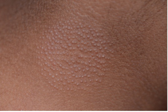
Fig. 2. The lesions are generally skin-colored, hyperkeratotic follicular papules that are 1 to 3 mm in diameter.
When the lesion is gently rubbed with a finger, it feels like a nutmeg grater.3 The horny spine of lichen spinulosus can be removed, leaving a tiny, funnel-like orifice in the papule.2,3 The papules often group or coalesce into larger patches or plaques (Figure 3) measuring 2 to 5 cm in diameter.5
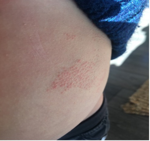
Fig. 3. The papules often group or coalesce into larger patches or plaques measuring 2 to 5 cm in diameter.
The distribution of the patches or plaques is often symmetrical.5,6 Sites of predilection include the trunk, extremities, neck, extensor surfaces of the arms (Figure 4), abdomen, buttocks, trochanters, and knees.2,3,13 Hands, face, and feet are usually spared.11

Fig. 4. Lichen spinulosus can be found on the trunk, extremities, neck, extensor surfaces of the arms, abdomen, buttocks, trochanters, and knees.
Occasionally, the lesions can be generalized, especially in those with an underlying disorder.4,10,11 The lesions are usually asymptomatic but may be mildly pruritic.6 The diagnosis is mainly clinical based on the typical features of patches or plaques of follicular papules topped by keratotic spines. Histological examination should be considered if the diagnosis is in doubt. The lesion of lichen spinulosus is confined to the skin. There is no known association with systemic disorders or genetic syndromes.4 The condition, however, can be cosmetically unsightly.5 The treatment course is variable. In most cases, lichen spinulosus remits spontaneously within 2 years,1,2 but lesions may persist for decades with a relapsing-remitting course.4,5
AFFILIATIONS:
1Clinical Professor of Pediatrics, the University of Calgary, Calgary, Alberta, Canada
2Pediatric Consultant, the Alberta Children’s Hospital, Calgary, Alberta, Canada
3Dermatologist, Medical Director and Founder, the Toronto Dermatology Centre, Toronto, Ontario, Canada
4Associate Clinical Professor of Pediatrics, Dermatology and Skin Sciences, the University of British Columbia, Vancouver, British Columbia, Canada.
5Pediatric Dermatologist, the Pediatric Institute, Kuala Lumpur General Hospital, Kuala Lumpur, Malaysia
CITATION:
Leung AKC, Barankin B, Lam JM, Leong KF. An atlas of lumps and bumps, part 39: lichen spinulosus. Consultant. 2024;64(5):e3. doi:10.25270/con.2024.05.000004
CORRESPONDENCE:
Alexander K. C. Leung, MD, #200, 233 16th Ave NW, Calgary, AB T2M 0H5, Canada (aleung@ucalgary.ca)
EDITOR’S NOTE:
This article is part of a series describing and differentiating dermatologic lumps and bumps. To access previously published articles in the series, visit: https://www.consultant360.com/resource-center/atlas-lumps-and-bumps.
REFERENCES
- Leung AKC, Barankin B. Flesh-colored lesions with a central horny spine: Lichen spinulosus. Consultant for Pediatricians. 2016;15:403-405
- Tilly JJ, Drolet BA, Esterly NB. Lichenoid eruptions in children. J Am Acad Dermatol. 2004;51(4):606-624. doi:10.1016/j.jaad.2003.12.012
- Friedman SJ. Lichen spinulosus. Clinicopathologic review of thirty-five cases. J Am Acad Dermatol. 1990;22(2 Pt 1):261-264.
- Venkatesh A, Dupuis E, Prajapati V, Rao J. Generalized lichen spinulosus in a 4-year-old boy without systemic disease. Arch Dermatol. 2012;148(7):865-866. doi:10.1001/archdermatol.2012.188
- Boyd AS. Lichen spinulosus: case report and overview. Cutis. 1989;43(6):557-560.
- Kim SH, Kang JH, Seo JK, Hwang SW, Sung HS, Lee D. Successful treatment of lichen spinulosus with topical tacalcitol cream. Pediatr Dermatol. 2010;27(5):546-547. doi:10.1111/j.1525-1470.2010.01275.x
- Uehara A, Abe M, Shimizu A, Motegi S, Amano H, Ishikawa O. Successful treatment of lichen spinulosus with topical adapalene. Eur J Dermatol. 2015;25(5):490-491. doi:10.1684/ejd.2015.2597
- Aghighi M, Pukhalskaya T, Brickley S, Smoller B. An uncommon case of lichen spinulosus in an adult patient clinically mimicking folliculotropic mycosis fungoides. Cureus. 2020;12(6):e8572. doi:10.7759/cureus.8572
- Lee ML, Piper DW, Fischer GO. Lichen spinulosus after the ingestion of omeprazole. Med J Aust. 1989;150(7):410. doi:10.5694/j.1326-5377.1989.tb136547.x
- Cohen SJ, Dicken CH. Generalized lichen spinulosus in an HIV-positive man. J Am Acad Dermatol. 1991;25(1):116-118. doi:10.1016/s0190-9622(08)80503-8
- Kabashima R, Sugita K, Kabashima K, Nakamura M, Tokura Y. Lichen spinulosus in an alcoholic patient. Acta Derm Venereol. 2009; 89(3):311-312. doi:10.2340/00015555-0600
- Kano Y, Orihara M, Yagita A, Shiohara T. Lichen spinulosus in a patient with Crohn's disease. Int J Dermatol. 1995;34(9):670-671. doi:10.1111/j.1365-4362.1995.tb01115.x
- Park JS. Lichen nitidus and lichen spinulosus or spinous follicular lichen nitidus? A second case. Clin Exp Dermatol. 2011;36(5):557-8. doi: 10.1111/j.1365-2230.2011.04026.x