Lumps and Bumps in Children: Abdominal and Inguinal Hernias
 Umbilical Hernia
The mass protruding from this 2-month-old boy’s umbilicus is an umbilical hernia. It was noted soon after the umbilical cord fell off and was soft. The mass enlarged when the infant cried or strained and was easily reducible inside the abdomen by external pressure.
An umbilical hernia results from imperfect closure or weakness of the umbilical ring. These hernias are common in infants and young children and make up roughly 5% to 10% of all primary hernias.1,2 Umbilical hernias are about 10 times more common in blacks than in whites.1 They are more common in low birth weight infants. The sex incidence is equal.2
Most umbilical hernias are sporadic and occur as isolated findings in otherwise healthy infants. Umbilical hernias occur with increased frequency in patients with Down syndrome, trisomy 13, trisomy 18, congenital hypothyroidism, Beckwith-Wiedemann syndrome, mucopolysaccharidosis, and cirrhosis of the liver with ascites.1
Classically, an umbilical hernia presents as a soft, skin-covered swelling that protrudes through the fibrous ring at the umbilicus. The umbilical bulge becomes more apparent during episodes of crying, coughing, or straining and is easily reducible. The content usually consists of a piece of small intestine and, sometimes, omentum. The condition is usually recognized in the neonatal period and is usually asymptomatic.1
Complications, such as incarceration of intestine or omentum, strangulation, perforation of the intestine, and rupture with evisceration, are rare in children.1,2 The risk of complications is much higher in adults.3 For young women with persistent umbilical hernia, the umbilical defect may enlarge and become symptomatic during pregnancy.1
Most umbilical hernias resolve spontaneously, usually within the first year of life.1 Rarely, surgery may become necessary if the hernia becomes incarcerated or strangulated, increases in size after the first year of life, or persists for 5 years.4 Repair at the age of 2 to 3 years is advocated by some surgeons when the fascial defect is greater than 1.5 cm in diameter, especially if the edge is thin and sharp.1,4,5 For those umbilical hernias with larger fascial defects, repair at a much earlier age is recommended.5 Of umbilical hernias not repaired in childhood, 10% persist into adulthood.5
REFERENCES:
1. Leung AK. Umbilical hernia. In: Leung AK, ed. Common Problems in Ambulatory Pediatrics: Specific Clinical Problems, volume 1. New York: Nova Science Publishers, Inc. (in press).
2. Sherman SC, Lee L. Strangulated umbilical hernia. J Emerg Med. 2004;26:209-211.
3. Ginsburg BY, Sharma AN. Spontaneous rupture of an umbilical hernia with evisceration. J Emerg Med. 2006;30:155-157.
4. Weik J, Moores D. An unusual case of umbilical hernia rupture with evisceration. J Pediatr Surg. 2005;40:E33-E35.
5. Durakbasa CU. Spontaneous rupture of an infantile umbilical hernia with intestinal evisceration.
Pediatr Surg Int. 2006;22:567-569.
 Paraumbilical Hernia
Unlike an umbilical hernia, a paraumbilical hernia does not protrude through the umbilical area. Rather, it protrudes just above or below the umbilicus. The paraumbilical hernia in this 7-year-old boy was first noted a year ago. It was reducible and asymptomatic.
The male to female ratio is about 1:5.1 The condition is more common in whites and obese persons.2 A paraumbilical hernia poses the risk of incarceration and strangulation.3,4 Omentum, small bowel, and large bowel are the usual content.1
The diagnosis of paraumbilical hernia can usually be made clinically, unless the hernia sac is small or the patient’s body habitus interferes with adequate palpation. High-resolution ultrasonography is an efficient tool for detecting the presence of a paraumbilical hernia and accurately verifies not only its content but also possible associated complications.3
A paraumbilical hernia does not close spontaneously.2 Elective herniorrhaphy is advisable because of the recognized risk of complications.
REFERENCES:
1. Daoud FS. Incarcerated endometriotic ovarian cyst within paraumbilical hernia. J Obstet Gynaecol. 2005;25:828-829.
2. Sinha SN, Keith T. Mesh plug repair for paraumbilical hernia. Surgeon. 2004;2:99-102.
3. Bedewi MA, El-Sharkawy MS, Al Boukai AA, Al-Nakshabandi N. Prevalence of adult paraumbilical hernia. Assessment by high-resolution sonography: a hospital-based study. Hernia. 2012;16:59-62.
4. Yau KK, Siu WT, Chan KL. Strangulated appendix epiploica in paraumbilical hernia: preoperative diagnosis and laparoscopic treatment. Surg Laparosc Endosc Percutan Tech. 2006;16:49-51.
 Epigastric Hernia
This abdominal mass in a 17-year-old boy appeared 3 years earlier and was asymptomatic. The boy was very active in sport activities. His past health was unremarkable. In particular, he had not undergone any surgery. On examination, the mass was not tender or reducible.
The mass is an epigastric hernia, which results from an intrinsic defect in the interstices of the decussating fibers of the linea alba.1 The hernia protrudes midline through the linea alba between the xyphoid and the umbilicus. It usually consists of extraperitoneal fat and, occasionally, a peritoneal sac that may contain abdominal viscera.2 Epigastric hernias account for 0.35% to 1.5% of all abdominal wall hernias.3,4 The condition is more common in males.5 The peak incidence is between 20 and 50 years of age.5 Physical straining has been suggested as a pathogenic factor.1
Although usually asymptomatic, an epigastric hernia may present with upper abdominal pain and dyspepsia.4 Epigastric hernia has to be differentiated from subcutaneous lipoma, fibroma, and neurofibroma.5 Sonography or CT scanning may help verify the diagnosis. Incarceration and strangulation are potential complications.1,4 Early surgical repair is therefore advocated.1
REFERENCES:
1. Lang B, Lau H, Lee F. Epigastric hernia and its etiology. Hernia. 2002;6:148-150.
2. Salameh JR. Primary and unusual abdominal wall hernias. Surg Clin North Am. 2008;88:45-60.
3. Asuquo ME, Nwagbara VI, Ifere MO. Epigastric hernia presenting as a giant abdominal interparietal hernia. Int J Surg Case Rep. 2011;2:243-245.
4. Cheung HY, Siu WT, Yau KK, et al. Incarcerated epigastric hernia, a rare cause of gastric outlet obstruction. J Gastrointest Surg. 2004;8:1111-1113.
5. Muschaweck U. Umbilical and epigastric hernia repair. Surg Clin North Am. 2003;83:1207-1221.
 Indirect Inguinal Hernia
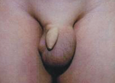
When this 6-year-old boy coughed or stood and performed the Valsalva maneuver, a mass appeared on the left side of his scrotum. The mass reduced spontaneously when he relaxed. The boy had no history of trauma to the area.
The hallmark of an indirect inguinal hernia is an intermittent bulge in the groin, scrotum, or labia that is most apparent with increased intra-abdominal pressure, such as with crying, straining, or coughing. An indirect inguinal hernia results from a failure of fusion of the processus vaginalis.1,2 The bowel subsequently descends through the inguinal canal and leads to hernia formation. The incidence of indirect inguinal hernia in term infants is 1% to 2%.2
About 50% of inguinal hernias present clinically in the first year of life, especially in the first 6 months.1,2 In premature infants, the incidence is 9% to 10%.2 The male to female ratio is 10:1.1,2 The incidence is higher in those infants with increased intra-abdominal pressure, undescended testis, congenital heart disease, cystic fibrosis, connective tissue disorders, bladder exstrophy, testicular feminization syndrome, and other intersex disorders.1,2 Inguinal hernia also occurs more commonly in patients with chromosomal disorders, microdeletion disorders, and single gene disorders.3 There is a familial tendency for hernia formation.1,2
The condition is often asymptomatic. Patients with incarcerated inguinal hernia may present with irritability, vomiting, abdominal distention, and a painful mass that is irreducible. The overlying skin may be erythematous. Incarceration occurs in about 12% of infants and young children with an inguinal hernia, often during the first 6 months of life.1,2 The rate of incarceration decreases to 1% at 8 years of age.4 Occasionally, strangulation may occur, and infarction of the small bowel, testis, or ovary may result.
The diagnosis is usually a clinical one. When the mass is not visually apparent, an older child can be asked to stand and perform a Valsalva maneuver, whereas a younger child may be allowed to cry to provoke an inguinal bulge to appear.2 The mass typically reduces spontaneously when the child relaxes or can be reduced by gentle pressure. The spermatic cord on the ipsilateral side is often thickened (silk string sign or silk glove sign).5 In the young girl, the hernia sac may contain the ovary and fallopian tube and present as a firm, discrete, nontender mass in the labia majora.
In patients with incarcerated inguinal hernia, plain radiographs may show multiple air-fluid levels. Ultra-sonography may help in the differentiation between a hernia and a hydrocele, if the clinical diagnosis is in doubt. A chromosome study should be ordered if testicular feminization syndrome is suspected.
Surgical repair of inguinal hernia shortly after diagnosis is recommended for all patients, including premature infants.6 If the child with an incarcerated hernia appears toxic or shows signs of peritonitis, manual reduction with the child sedated can be attempted. Immediate surgery is indicated for the incarcerated hernia that is manually irreducible. Complications of inguinal hernia surgery include wound infection, hematoma, scrotal edema, ascent of testis, testicular atrophy, and recurrence.4
There is controversy about whether the contralateral groin should be explored.7 Today, most surgeons do not routinely perform a contralateral exploration unless a contralateral inguinal hernia or patent processus vaginalis can be demonstrated either by preoperative ultrasonography or intraoperative laparoscopy.7
Laparoscopic inguinal hernia repair has become an alternative to the conventional herniotomy/open procedure.7 This procedure is safe, reproducible, and technically easy for experienced laparoscopic surgeons.7 It also does not impair testicular perfusion.7 The main advantages of laparoscopic inguinal hernia repair over conventional herniotomy are less pain, earlier recovery, better wound cosmesis, and the ability to detect and simultaneously repair contralateral patency of processus vaginalis.8
REFERENCES:
1. Leung AK. Indirect inguinal hernia. In: Leung AK, ed. Common Problems in Ambulatory Pediatrics: Specific Clinical Problems, volume 1. New York: Nova Science Publishers, Inc. In press.
2. Leung AK. Indirect inguinal hernia. In: Lang F, ed. The Encyclopedia of Molecular Mechanism of Disease. Berlin: Springer-Verlag; 2009:834-835.
3. Barnett C, Langer JC, Hinek A, et al. Looking past the lump: genetic aspects of inguinal hernia in children. J Pediatr Surg. 2009;44:1423-1431.
4. Rosenberg J. Pediatric inguinal hernia repair—a critical appraisal. Hernia. 2008;12:113-115.
5. Luo CC, Chao HC. Prevention of unnecessary contralateral exploration using the silk glove sign (SGS) in pediatric patients with unilateral inguinal hernia. Eur J Pediatr. 2007;166:667-669.
6. Vaos G, Gardikis S, Kambouri K, et al. Optimal timing for repair of an inguinal hernia in premature infants. Pediatr Surg Int. 2010;26:379-385.
7. Lau ST, Lee YH, Caty MG. Current management of hernias and hydroceles. Semin Pediatr Surg. 2007;16:50-57.
8. Chan KL, Chan HY, Tam PK. Towards a near-zero recurrence rate in laparoscopic inguinal hernia repair for pediatric patients of all ages. J Pediatr Surg. 2007;42:1993-1997.


