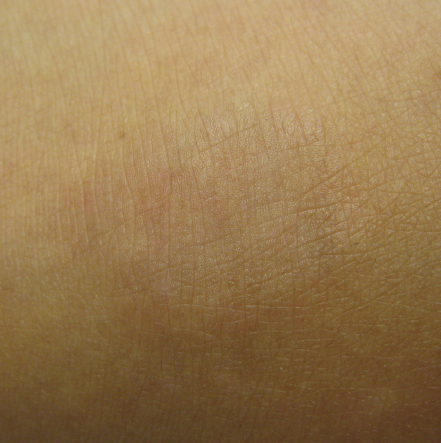
A Circular Lesion on the Back of the Foot
A 5-year-old African American girl presented with a skin lesion on the dorsum of her left foot. The lesion had been present for 2 to 3 months. She reported that it had started as a small bump and gradually had enlarged, taking on a circular shape with a raised edge and normal-appearing skin in the center. The lesion was not painful or pruritic.


What do you suspect is causing this lesion?
(Answer and discussion on next page)
Answer: Granuloma Annulare
Localized GA usually presents as small papules that gradually form a raised edge of an enlarging circle. It usually is violaceous in the white population or skin-colored in people with darker skin. The absence of scales can help in differentiating GA from other circular skin lesions (mainly tinea corporis) and the herald patch of pityriasis rosea. The most common affected sites are the dorsum of the hand or foot, but GA can affect any part of the body.
The diagnosis is clinical; biopsy is rarely needed to diagnose localized GA. Histologically, GA is characterized by the presence of focal degeneration of collagen and deposition of mucin and lymphocytic infiltration in the upper and middle dermis.
GA is a self-limiting condition, usually resolving spontaneously within a few months to 2 years. Topical corticosteroids have been shown to shorten the duration of GA1; topical tacrolimus also might be effective.2
Hytham Fadl, MD, is a pediatrician at Hurley Medical Center in Flint, Michigan.
References
1. Rallis E, Stavropoulou E, Korfitis C. Granuloma annulare of childhood successfully treated with potent topical corticosteroids previously unresponsive to tacrolimus ointment 0.1%: report of three cases. Clin Exp Dermatol. 2009;34(7):e475-e476.
2. Harth W, Linse R. Topical tacrolimus in granuloma annulare and necrobiosis lipoidica. Br J Dermatol. 2004;150(4):792-794.


