ABSTRACT: Chronic recurrent multifocal osteomyelitis (CRMO) is an inflammatory bone disease that occurs primarily in childhood. The clinical picture often is confused with bacterial osteomyelitis. Awareness of CRMO as a clinical entity helps avoid diagnosis and treatment delays. Our patient, an 8-year-old girl, presented with acute left hip pain. One month after presentation, a lytic lesion was seen on plain radiographs; biopsy revealed nonspecific inflammation. It was not until more than 2 years later, when multifocal bone lesions and psoriasis developed, that the diagnosis became clear. Our patient’s case demonstrates several key points: not all children with CRMO present with multifocal disease, patients frequently have comorbid inflammatory conditions, and there are no diagnostic laboratory studies. The optimal treatments remain unknown.
Chronic recurrent multifocal osteomyelitis (CRMO) is a rare chronic inflammatory bone disease that occurs mostly in children.1,2 Patients present with bone pain that may be accompanied by fever. CRMO often is associated with inflammatory disorders of the skin and GI tract.
Plain radiographs demonstrate osteolytic lesions surrounded by sclerosis; lesions usually are located in the metaphyses of the long bones.3 The erythrocyte sedimentation rate (ESR) and C-reactive protein (CRP) level often are mildly elevated.2,4,5 There are no diagnostic laboratory studies.
The clinical picture of CRMO may be confused--most often with bacterial osteomyelitis. Biopsy results are consistent with osteomyelitis, but culture results are nearly always normal.1,2,4
A lack of awareness of CRMO as a clinical entity contributes to delayed diagnosis, which often leads to unnecessary patient exposure to antibiotics, repeated biopsies, prolonged hospitalization, and delayed institution of appropriate treatment. In this brief review, we offer an illustrative case report, describe key features that help establish the diagnosis, and discuss the treatment options and outcome.
CASE REPORT
An 8-year-old white girl presented with acute left hip pain without associated fever. Plain x-ray films of the hip were ordered; the results were normal. A diagnosis of toxic synovitis was made. The girl's symptoms improved but recurred 10 days later.
A bone scan revealed uptake in the girl's left proximal femur. MRI findings were consistent with bone marrow edema in the left greater trochanter. The girl remained afebrile and had a normal complete blood cell (CBC) count and ESR; therefore, observation without antibiotics was recommended.
The girl continued to have daily left hip pain. Repeated radiographs of her left hip 4 weeks after the onset of symptoms revealed a radiolucent area in the left greater trochanter, prompting a more extensive workup. The girl's ESR was 35 mm/h, and her CBC count was normal.
A bone biopsy specimen showed stromal edema, trilineage hemato- poiesis, focal acute hemorrhage, and hemosiderin-laden macrophages. No granulomas or malignant cells were seen. The results of CD1a staining were negative, ruling out Langerhans cell histiocytosis. Cultures of the bone and blood were sterile.
The girl was treated with ibuprofen(, but her pain became so severe that she spent 1 month in a wheelchair. However, 2 months after treatment was started, the girl was nearly asymptomatic.
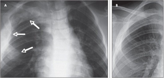
Scapular pain in cycles. At age 10 years, the girl experienced severe right scapular pain that recurred in roughly 9-week cycles. Shortly after the onset of scapular pain, an erythematous scaly rash developed on her anterior chest overlying the right third rib. A chest x-ray film revealed lytic bone lesions in the right third rib (Figure 1).
Figure 1 – This chest radiograph obtained for our patient with chronic recurrent multifocal osteomyelitis (A) demonstrates bony enlargement and extensive osteolytic lesions with areas of sclerosis in her right third rib (arrows). Marked improvement was seen in the third rib after the patient was treated with methotrexate (B).
A bone scan obtained 6 months later demonstrated increased uptake on delayed images in the right third rib (Figure 2), the greater trochanter of the left femur, and the right first metatarsal. Biopsy specimens of the third rib revealed osteoblastic rimming of the bony trabeculae and paratrabecular fibrosis with scattered collections of plasma cells and lymphoid cells; there was no evidence of malignancy. The results of CD1a and S100 staining were normal, ruling out Langerhans cell histiocytosis.
Figure 2 – A technitium99 bone scan obtained for this patient shows increased radiotracer
uptake diffusely in the right third rib on delayed images.
A punch biopsy of the skin lesion displayed epidermal hyperke- ratosis, parakeratosis, and acanthosis. There were small subcorneal pustules and patchy neutrophilic infiltrate within the dermis and patchy perivascular lymphocytic infiltrate in the superficial dermis, consistent with psoriasis.
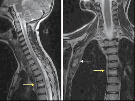
Scoliosis at 11 years. At age 11 years, the girl continued to have rib and scapular pain and high-left thoracic scoliosis developed. Her CBC count and CRP level were normal; her ESR was 57 mm/h. The results of an antinuclear antibody test were normal. A CT scan of the thoracic spine revealed partial volume loss with faint sclerosis in the upper half of the T6 vertebral body. MRI dem- onstrated increased uptake in the T6 vertebra (Figure 3). The girl's pain became persistent.
Figure 3 – An MRI demonstrates partial volume loss of the upper aspect of the T6 vertebral body on the right (yellow arrows) in our patient with chronic recurrent multifocal osteomyelitis. In addition, the third rib (white arrow) is enlarged and has scattered areas of increased signal intensity.
Just before the girl's 12th birthday, left foot pain developed. Plain x-ray films revealed bony changes in the proximal phalanx of the fourth and fifth toes. Then, right-sided mandibular pain developed and the girl underwent surgery to remove tissue overlying an unerupted molar.
Several weeks later, marked swelling of the girl's right mandible developed. Examination revealed a firm right mandibular mass. Radiographs of the mandible demonstrated radiolucency with surrounding sclerosis. The biopsy revealed immature and mature bone with fibrosis; there was chronic inflammation of the medullary space that was consistent with chronic osteomyelitis but no evidence of malignancy.
Culture of the bone (obtained via an intra-oral route) grew mixed oral flora. Because the culture result was abnormal, the girl was treated with clindamycin(. However, the symptoms worsened. After several weeks, she was switched to azithromycin(, which was associated with improvement of her mandibular pain and swelling; however, the pain in her scapula, knee, and toes was undiminished.
Daily pain and ongoing erythema. When the girl presented to our clinic, she was 12 years and 4 months old. She complained of daily pain in the right lateral distal femur, right scapula, left fourth and fifth metatarsophalangeal joints, and right man- dible. She continued to have frequent plaques of erythema covered with a silver scale on the skin of her trunk, feet, and scalp. She complained of daily bone pain, which she rated as 4 to 7 on a pain scale of 10, even though she was taking ibuprofen.
The girl's past medical history was remarkable for dysautonomia; short stature (height below the third percentile), accompanied by a 2.5-year delayed bone age; and problems with pustular psoriasis on her feet. Her family history was significant for psoriasis in her mother and maternal grandfather. Her maternal uncle had psoriasis and psoriatic arthritis. Her paternal grandmother had rheumatoid arthritis (RA). Her two younger siblings were in good health. The review of systems showed frequent aphthous ulcers.
On physical examination, the girl was afebrile; her height and weight were below the third percentile. She had several small, shallow oral ulcers on the buccal mucosa. She had mild pain on palpation over the right third rib, the lateral aspect of the left distal femur, the distal left metatarsal heads of the fourth and fifth toes, and the right scapula. There was a hard, nonmobile mass on the right mandible. The girl had palpable bony enlargement of the right third rib. She also had erythematous plaques with overlying silver scale on her feet and the extensor surfaces of her knees, abdomen, and scalp. Examination of the nails revealed multiple nail pits. The rest of the examination results were unremarkable.
Laboratory studies revealed a normal CBC count, ESR, and CRP level. The biopsies and radiographs were reviewed and a diagnosis of CRMO was confirmed.
Improvement seen with treatment. Multiple treatment options were discussed with the patient and her family, including the use of corticosteroids, methotrexate( (MTX), tumor necrosis factor a (TNF-a) inhibitors, and bisphosphonates. They opted for a trial of MTX. The patient started taking 20 mg of MTX orally once each week (0.75 mg/kg/wk) with daily folic acid( supplementation.
The girl showed marked improvement in her bone pain and resolution of her mandibular swelling. Her energy level returned to normal; she had been missing school but was no longer, and her grades improved dramatically. She has had no major disease exacerbations for 1 year since starting MTX. She continues to have intermittent short-lived episodes of mild bone pain, but they do not interfere with her activities.
KEY DIAGNOSTIC FEATURES
Disease spectrum versus same disease with lots of acronyms. Our patient's case illustrates several important points about CRMO. Not all children with CRMO present with multifocal disease. Our patient presented with unifocal disease, and multifocal lesions did not develop until 2 years later. With only uni- focal disease, she would not have met the criteria for CRMO that Björkstén and Boquist6 proposed, which include multifocal bone lesions, prolonged clinical course over several years, recurrent epi- sodes of bone inflammation, and lack of response to prolonged anti- microbial therapy. Other children may have unifocal recurrent disease but never additional lesions; they have a similar clinical, radiographic, and histological picture but would not fall under the diagnostic category of CRMO because of the lack of multifocal lesions.4,5
Girschick and colleagues5 suggested that the term "chronic nonbacterial osteomyelitis" (CNO) be used instead of CRMO so that the spectrum of disease would be covered by 1 diagnostic label. Other names for the same disorder include nonbacterial osteitis (NBO), symmetric osteomyelitis, pustulotic ar- thro-osteitis, chronic sclerosing osteomyelitis of Garré, and lymphoplasmacellular osteomyelitis.
To further complicate things, many children with CRMO often receive a diagnosis of synovitis, acne, pustulosis, hyperostosis, osteitis (SAPHO) syndrome--especially children who have CRMO and palmoplantar pustulosis. Although classic SAPHO and CRMO have clinical differences, they are quite similar and probably are part of the same disease spectrum.
Jansson and associates4 recently proposed diagnostic criteria for the CRMO/CNO/NBO disease spectrum that are more inclusive than the original Björkstén and Boquist criteria. However, the newer criteria need to be validated independently. Standardizing the nomenclature and having clinically validated diagnostic criteria would be beneficial.
Comorbid inflammatory conditions are common. Patients with CRMO frequently have comorbid inflammatory conditions. The usual involvement of the skin and the GI tract suggests that a common inflammatory pathway is shared in the bone, skin, and gut.
The most frequently seen comorbid conditions include palmoplantar pustulosis and psoriasis vulgaris, both of which our patient had during her disease course.2,7,8 Other associated inflammatory disorders include Crohn disease, ulcerative colitis, pyoderma gangrenosum, arthritis, vasculitis, and sacroiliitis.1,2,4,5,8,9 In addition, early-onset CRMO is associated with a congenital dyserythropoietic anemia and Sweet syndrome in Majeed syndrome, a rare autosomal recessive disorder.10
A possible genetic component. A family history of chronic inflammatory disorders suggests a genetic component to disease. Our patient had 4 family members (first- and second-degree relatives) who had psoriasis or RA. Nearly half the families in 1 cohort had a first- or second-degree relative who had a chronic inflammatory disorder, most often a form of psoriasis4; in that cohort, 6% of the families had more than 1 member with nonbacterial osteitis. Further evidence for a genetic basis of the disease includes identification of a susceptibility locus for sporadic CRMO on chromosome 18, identification of the gene for Majeed syndrome as LPIN2, and mutations in pstpip2 found in 2 mouse models of the disease.11-14
Remarkably normal findings. Physical examination and laboratory study results are remarkably normal. When our patient presented initially with a unifocal bone lesion, she did not appear to be ill. She had an unremarkable physical examination, except for the finding of pain over the site of the bone lesion. Her laboratory study results were unremarkable, including a normal CBC count and ESR, and 1 month later, her ESR was moderately elevated. In a cohort of 39 patients who had a multifocal disease course or unifocal disease plus palmoplantar pustulosis, 85% had an elevated ESR, 79% had an elevated CRP level, and 66% had a TNF-a level higher than 25 pg/mL.4
Benefits with radiographic studies. Radiographic studies are useful in making the diagnosis of CRMO. Characteristic changes on plain x-ray films include lytic lesions with surrounding sclerosis in the metaphyses of the long bones adjacent to the growth plate. A mild periosteal reaction also may be present and later, the long bone lesions show evidence of sclerosis; over time, the osseous structure may return to normal.3 In contrast, clavicular lesions typically demonstrate a significant periosteal reaction accompanied by lytic destruction of the medullary bone; significant sclerosis and hyperostosis often occur later.
Bone scanning is an important diagnostic tool because many lesions are clinically silent; however, they may be detected by scintigraphy.3,8 MRI with short tau inversion recovery (STIR) images helps clinicians determine the extent of disease and disease activity. Classically, an active lesion demonstrates increased signal intensity on STIR and T2-weighted images and decreased signal intensity on T1-weighted images.3
Diagnosis helped by bone biopsy. A bone biopsy is invaluable in establishing a definitive diagnosis. It serves at least 3 purposes: (1) provides a source of tissue for culture to rule out infectious osteomyelitis; (2) eliminates leukemia, lymphoma, Ewing sarcoma, histiocytosis, and eosinophilic granuloma as diagnostic possibilities; and (3) documents inflammation consistent with the diagnosis of CRMO.
Typical histological features of CRMO early in the disease course include an inflammatory infiltrate that has a predominance of neutrophils.6 Additional features that may be present include necrotic fragments, osteoclastic bone resorption, multinucleated giant cells, granulomatous foci, and abscess formation. Later, a lymphocytic infiltrate with areas of fibrosis and new bone formation may be seen.
TREATMENT AND OUTCOME
Treatment is empiric. The optimal treatment strategies for patients with CRMO remain unknown. Interpreting response to treatment becomes a challenge because of the rare nature of the disorder, complicated by a disease course that includes spontaneous remissions.
NSAIDs typically are chosen as the first line of treatment; they provide symptomatic relief in up to 80% of patients.2,4,5,9 However, as many as 50% of patients continue to have symptoms despite NSAID therapy and require other medications.5
Corticosteroids often are the second-line agents used to manage CRMO; they provide at least transient pain relief in most patients.2,4,5,7,9 However, the unfavorable adverse effect profile of corticosteroids makes them a poor choice for long-term treatment. MTX and sulfasalazine( have been used in some patients; success has been reported, and generally they are well tolerated.5,9 However, some children do not respond to these drugs and more aggressive therapy is needed.4
Dramatic improvement has been reported after treatment with TNF-a inhibitors and with intravenous bisphosphonates.4,15,16 Again, however, not all patients respond. Improvement in small numbers of patients has been reported with interferon-a; interfer- on-g; colchicine(; hyperbaric oxygen; azithromycin (thought to be beneficial because of its anti-inflammatory effect rather than its antimicrobial effect); and curettage or partial resection,2,17-19 although not in all cases.2,4,5 Surgical intervention usually is limited to obtaining a biopsy for culture and histological evaluation.
Outcome generally good. For most patients with CRMO, the long-term outcome is good. However, most experience significant morbidity during periods of active disease. The average duration of disease activity in cohorts of Canadian/Australian and French children with CRMO was 5.6 years and 5.3 years, respectively.1,9 In the Canadian/Australian population, 26% of participants continued to have active disease more than 9 years after diagnosis; 57% of those in the French cohort had active disease after a mean follow-up of 5.3 ± 2.5 years.1,9
Significant bony deformities that the participants considered either functionally or cosmetically problematic developed in as many as 50%.1 School performance, job performance, and job choices were affected adversely in some of the patients. Chronic pain that generally was associated with continued disease activity was reported in a subset of patients. Participants who had comorbid inflammatory conditions often had ongoing problems with active inflammatory disease--including arthritis, psoriasis, and inflammatory bowel disease--long after the bone lesions had become asymptomatic.
SUMMARY
Because the clinical picture of CRMO often is confused and there are no diagnostic laboratory studies, radiographic evaluation and bone biopsy are useful in making the diagnosis. Treatment is empiric, but anti-inflammatory agents often are used and purportedly are successful. Although the long-term outcome is good for most patients, significant morbidity occurs during periods of disease activity. Early recognition and management of CRMO probably will reduce the associated short- and long-term morbidity. *


