Bacterial Meningitis in Children: Could It Be Mondini Dysplasia?
Bacterial meningitis in children is uncommon in the post-Haemophilus influenzae type b (Hib) and pneumococcal conjugate vaccine era.1,2 The occurrence of recurrent bacterial meningitis is so rare that investigations for anatomical defects are routinely performed. Mondini defects are a rare type of congenital inner ear abnormality of which the clinician may not be aware. Here we provide a case and brief review of Mondini dysplasia and its association with meningitis.
CASE: A previously healthy, fully vaccinated 4-year-old girl was hospitalized for bacterial meningitis. Her recent medical history was significant for clear discharge from her left ear for the past 3 months. Her past medical history was significant for deafness in her left ear discovered during an evaluation for speech delay.
On physical examination, the patient was febrile. Her left ear canal was draining serous fluid. Her neurological examination was remarkable for lethargy, irritability, and slurred speech. An axial CT scan of the head was normal except for the finding of fluid in the left mastoid air cells. A Gram stain of her cerebrospinal fluid (CSF) showed 41 gram-positive cocci in pairs and chains; the culture grew Streptococcus pneumoniae serotype 19A.

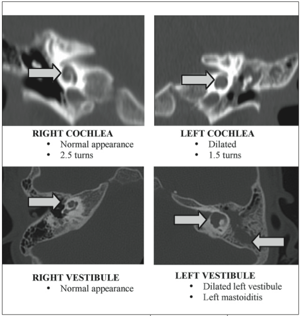
Figure – A high-resolution CT scan of the temporal bones highlight the normal right cochlea and vestibule compared to the classic findings of Mondini dysplasia in the left cochlea and vestibule (with associated mastoiditis).
The hospital course was complicated by seizure activity and left facial nerve paralysis. A high-resolution CT scan of the temporal bones demonstrated the Mondini defect (Figure).
The patient was treated with intravenous antibiotics for 10 days and, upon discharge, received the 23-valent pneumococcal polysaccharide and 4-valent conjugate meningococcal vaccines. In the following month, the patient underwent definitive repair of the Mondini defect.
PREDISPOSING FACTORS OF BACTERIAL MENINGITIS
Since the introduction of the Hib and S pneumoniae conjugate vaccines in 1990 and 2000, respectively, bacterial meningitis in the United States has become an infection seen more commonly in adults than in infants and young children.1 While S pneumoniae remains the most frequent cause of bacterial meningitis, most pediatric cases are caused by nonvaccine serotypes.2
Recurrent bacterial meningitis is so unusual that investigation for an underlying predisposing condition should be undertaken in any child with more than one episode of meningitis. Further evaluations should include appropriate imaging studies to detect the presence of anatomic defects as well as immunologic studies to determine whether the patient has an underlying immunodeficiency.3,4
The imaging study of choice is a high-resolution CT scan of the temporal bones. A routine head CT scan is of insufficient resolution to define the cochlear anatomy. Included among the anatomic defects that can predispose a patient to the development of bacterial meningitis are congenital abnormalities of the inner ear, including Mondini dysplasia.
MONDINI DYSPLASIA: CLINICAL FEATURES AND EVALUATION
The original 1791 scientific description of this interesting anatomic malformation is entitled “The anatomical section of a boy born deaf.”5 In this report, Carlo Mondini described the 3 main features of the inner ear abnormalities that now carry his name:
•A cochlea of 1.5 turns instead of the normal 2.5 turns, comprising a normal basal turn and a cystic apex in place of the distal 1.5 turns.
•An enlarged vestibule with normal semicircular canals.
•An enlarged vestibular aqueduct containing a dilated endolymphatic sac.
Usually, Mondini dysplasia is associated with some degree of hearing impairment and can be associated with CSF otorrhea and bacterial meningitis.6,7 It is thought that a fistula between the CSF spaces and the middle ear allows bacterial seeding of the meninges. The fistula itself can occur as a deficient oval window or stapes footplate.
In patients suspected to have Mondini dysplasia or who present with recurrent bacterial meningitis, the medical history and physical examination should include a review of CSF otorrhea and/or rhinorrhea, head trauma, previous surgeries of the ears, sinuses, skull base, and hearing impairment or speech delay. Most affected patients have profound sensorineural hearing loss, but hearing may be preserved.6-8
IMPORTANCE OF IMMUNIZATION
It is important to immunize patients with Mondini dysplasia to prevent future episodes of bacterial meningitis. Conjugate pneumococcal and Hib vaccines are routine during infancy (make sure the vaccines are up-to-date!), and the 23-valent pneumococcal polysaccharide vaccine is indicated for all children 2 years and older with anatomic inner ear abnormalities and in patients with cochlear implants.9
Our patient was not infected with pneumococcal serotypes included in the 7-valent pneumococcal conjugate vaccine. Since 2010, the 7-valent pneumococcal conjugate vaccine has been replaced by the 13-valent pneumococcal conjugate vaccine.
KEY POINTS FOR YOUR PRACTICE
Mondini dysplasia should be suspected in any child with hearing loss and bacterial meningitis because most patients with Mondini dysplasia have some degree of baseline hearing loss. We perform high-resolution CT or MRI of the temporal bones to optimize visualization of the inner ear anatomy in all patients with recurrent bacterial meningitis and in any of our patients with bacterial meningitis when there is a history of otorrhea or known sensorineural hearing loss. Early identification of an anatomic defect, such as Mondini dysplasia with a CSF communication, is necessary to prevent recurrent meningitis using recommended immunizations and appropriate surgical intervention.


