A Gallery of Pigmentation Disorders

Oral Hyperpigmentation: Two Cases
Gingival hyperpigmentation was noted in a 27-year-old African American man during a routine physical examination (A). It had been present since birth, and the patient was unaware of similar hyperpigmentation in other family members.
In another case, a 28-year-old African American man had had two patches of hyperpigmentation on the dorsum of the tongue since birth (B). He had no other areas of hyperpigmentation. His mother and several relatives on her side of the family were born with similar oral hyperpigmented areas.
Among African Americans and others who are dark-skinned, oral hyperpigmentation is usually physiologic. The most common sites are the gingiva and the buccal mucosa, although hyperpigmented areas may develop anywhere in the oral cavity.

These melanin deposits are of no clinical consequence. If the patient wishes to have them removed for cosmetic reasons, chemical peeling with phenol may be used. This destroys the melanin in the germinative layer of the epithelium.1
REFERENCE:
1. Lawson W. Pigmented oral lesions: clues to identifying the potentially malignant. Consultant. 2012;52(5):347-352.
 Congenital Hypopigmentation
This area of hypopigmentation had been present on the left buttock of a healthy 4-year-old African American girl since birth. The lesion is nevus depigmentosus, which is sometimes referred to as nevus achromicus. It occurs in about 1 in 50 to 75 persons in the general population.1 Nevus depigmentosus is usually present at birth as a solitary hypopigmented patch. This patient’s lesion is larger than most, which typically measure a few centimeters in diameter and have irregular but well-defined borders. In contrast to the lesions of vitiligo, which contain no pigmentation, the patches of nevus depigmentosus are hypopigmented. Nevus depigmentosus requires no treatment.
REFERENCE:
1. Ortonne JP. Vitiligo and other disorders of hypopigmentation. In: Bolognia JL, Jorizzo JL, Rapini RP. Dermatology. St Louis: Mosby; 2003:947-973.
 Postinflammatory Hypopigmentation and Hyperpigmentation
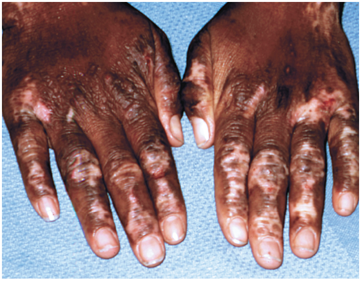
Scattered areas of irregular hypopigmentation and, to a lesser extent, hyperpigmentation (over some of the knuckles) were noted on the dorsal surfaces of the hands of a 28-year-old African American woman during a routine physical examination. She reported that the areas of abnormal pigmentation had developed 2 years earlier.
The patient had a 20-year history of eczema with inflammatory reaction, which involved the hands and antecubital and popliteal areas. The condition worsened when she experienced stress.
Inflammation from allergic reactions, infection, or trauma can result in either hypopigmentation or hyperpigmentation of the involved area. Although postinflammatory hypopigmentation after an episode of eczema is unusual, it may be more common—or more noticeable—in persons with dark-pigmented skin.1
This patient was treated with tacrolimus ointment, triamcinolone cream, fluocinonide cream, and oral prednisone.
REFERENCE:
1. Habif TP. Clinical Dermatology: A Color Guide to Diagnosis and Treatment. 4th ed. St Louis: Mosby; 2004:692.
 Vitiligo: Two Cases
A 56-year-old African American woman presented with perianal and, to a lesser degree, vulvar hypopigmentation (A). It was asymptomatic. She shaved the area every 2 weeks and had noticed the change in color about 6 weeks earlier. She had no known family members with vitiligo.
In another case, a 31-year-old white woman reported that vitiligo of the lower vulvar and perianal areas had developed after childbirth 4 years earlier (B). The condition was asymptomatic.
Vitiligo results from the complete absence of melanocytes in the epidermis. The condition affects approximately 1% of the general population; 50% of cases occur before 20 years of age.1 There is no sex predilection. Common sites are the face, neck, dorsal surfaces of the hands, and body folds, including axillae and genitalia.1
 The suspected cause of the depigmentation is an autoimmune event in persons with an underlying genetic predisposition. Those who are affected may have a family history of autoimmune disease or a family member with vitiligo.
Repigmentation may occur naturally or after treatment; however, pigment loss may occur all over the body and progress despite treatment. Vitiligo is most commonly treated with topical corticosteroids. For patients with small areas of depigmentation, topical immunomodulators and photochemotherapy have also been effective. When vitiligo is extensive, depigmentation of the remaining pigmented skin is an option.
REFERENCE:
1. Habif TP. Clinical Dermatology: A Color Guide to Diagnosis and Treatment. 4th ed. St Louis: Mosby; 2004:684-689.


